![]()
ITM University Gwalior is ranked among the best as Platinum Category Engineering Institute in India by AICTE, Ministry of HRD, Govt. of India in 2017.
T: 1800 270 0031
Email: admissions@itmuniversity.ac.in
ITM University
NH-44,BypassTurari, Jhansi Road Gwl (M.P.) 475001,(INDIA)
-
About
About ITM University
ITM University Top-Ranked Education
 Leadership
 Recognitions
& Approvals What Gwalior Offers
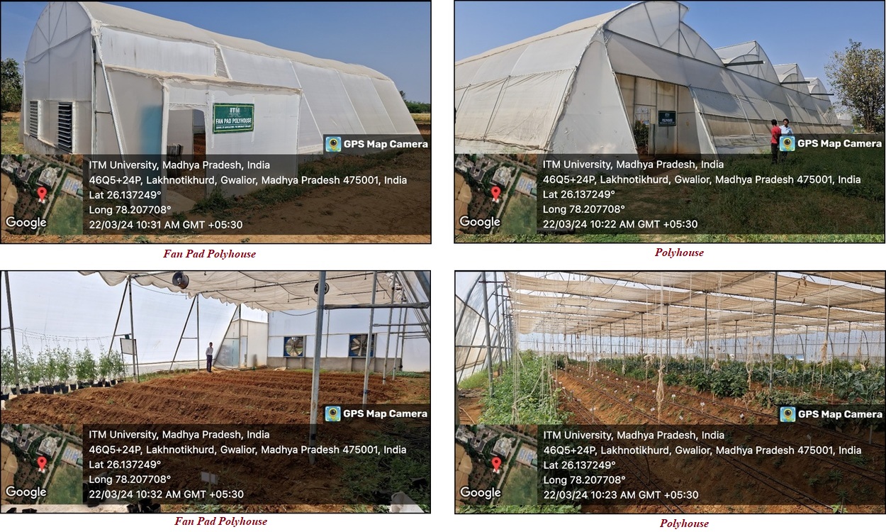
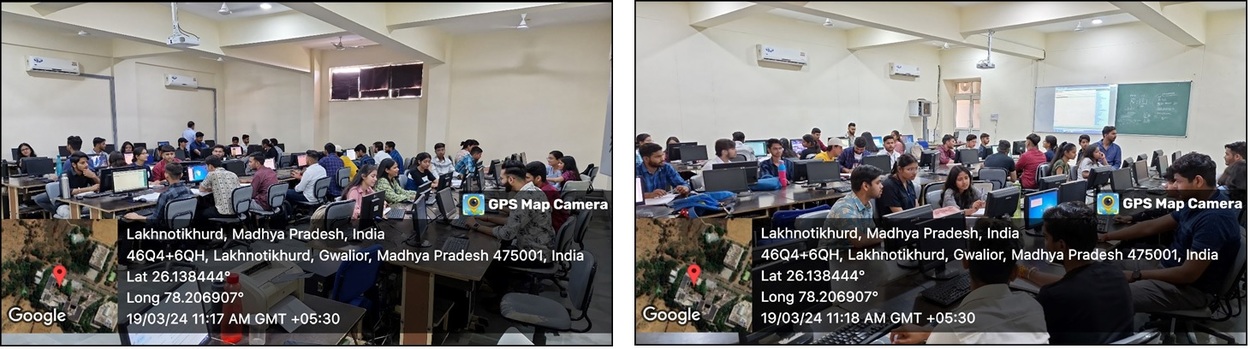
 Infrastructure
-
Admissions
Realize Your Professional Dreams
 Eligibility Criteria
 Fee Structure (2025-26)
 Scholarship Policy
 Ph.D. Notifications
-
Placements
Connecting Talent with Opportunity
Dark Mode